Labounek, Renรฉ, Bondy, Monica T., Paulson, Amy L., Bรฉdard, Sandrine, Abramovic, Mihael, Alonso-Ortiz, Eva, Atcheson, Nicole T., Barlow, Laura R., Barry, Robert L., Barth, Markus, Battiston, Marco, Bรผchel, Christian, Budde, Matthew D., Callot, Virginie, Combes, Anna, De Leener, Benjamin, Descoteaux, Maxime, de Sousa, Paulo Loureiro, Dostรกl, Marek, Doyon, Julien, Dvorak, Adam V., Eippert, Falk, Epperson, Karla R., Epperson, Kevin S., Freund, Patrick, Finsterbusch, Jรผrgen, Foias, Alexandru, Fratini, Michela, Fukunaga, Issei, Wheeler-Kingshott, Claudia A.M. Gandini, Germani, GianCarlo, Gilbert, Guillaume, Giove, Federico, Grussu, Francesco, Hagiwara, Akifumi, Henry, Pierre-Gilles, Horรกk, Tomรกลก, Hori, Masaaki, Joers, James M., Kamiya, Kouhei, Karbasforoushan, Haleh, Keลkovskรฝ, Miloลก, Khatibi, Ali, Kim, Joo-Won, Kinany, Nawal, Kitzler, Hagen, Kolind, Shannon, Kong, Yazhuo, Kudliฤka, Petr, Kuntke, Paul, Kurniawan, Nyoman D., Kusmia, Slawomir, Laganร , Maria Marcella, Laule, Cornelia, Law, Christine S.W., Leutritz, Tobias, Liu, Yaou, Llufriu, Sara, Mackey, Sean, Martin, Allan R., Martinez-Heras, Eloy, Mattera, Loan, OโGrady, Kristin P., Papinutto, Nico, Papp, Daniel, Pareto, Deborah, Parrish, Todd B., Pichiecchio, Anna, Prados, Ferran, Rovira, รlex, Ruitenberg, Marc J., Samson, Rebecca S., Savini, Giovanni, Seif, Maryam, Seifert, Alan C., Smith, Alex K., Smith, Seth A., Smith, Zachary A., Solana, Elisabeth, Suzuki, Yuichi, Tackley, George W, Tinnermann, Alexandra, Valoลกek, Jan, Van De Ville, Dimitri, Yiannakas, Marios C., Weber II, Kenneth A., Weiskopf, Nikolaus, Wise, Richard G., Wyss, Patrik O., Xu, Junqian, Cohen-Adad, Julien, Lenglet, Christophe, & Nestraลกil, Igor. (2025). *Imaging Neuroscience, 3*, imag_a_00559.
Clinical research aims to use careful and consistent study designs, often matching groups by factors like age and sex to reduce biological differences. However, many studies donโt take into account body sizeโsuch as height and weightโwhich could affect results, especially in brain and spinal cord imaging. This study explores how body size is related to measurements taken from MRI scans of the brain and spinal cord.
We analyzed data from 267 healthy adults (average age about 30 years, including 125 women) from a diverse population. The results show that body height is linked to various brain and spinal cord structures. Taller individuals tend to have larger volumes of brain gray matter, cortical gray matter, cerebellum, brainstem, and a larger cross-sectional area of white matter in the cervical spinal cord (correlation values ranging from 0.44 to 0.62). Intracranial volume (ICV)โthe total space inside the skullโalso correlates with height (r = 0.46) and with these brain and spinal cord measures (ranging from 0.37 to 0.77).
By comparison, age had weaker relationships, mainly with gray matter and cortical thickness (r values between -0.21 and -0.27). Body weight also showed specific associations, especially with spinal cord tissue propertiesโsuch as magnetization transfer ratio and diffusivityโbut mostly in men.
Measurements of spinal cord white matter were related to brain volumes and certain brain tissue properties. Statistical models showed that using only age and sex could explain 2% to 26% of differences in brain and spinal cord size. But when body height and ICV were added, this increased to 33% to 46%. In women, these explanations were about half as strong, suggesting that other unknown biological factors may influence the structure of the central nervous system in females.
In conclusion, body size and intracranial volume are important biological variables. Along with sex and age, body size should be included in the design and analysis of clinical studies that examine brain and spinal cord structure. Adjusting brain measurements by ICV reduces the effects of body size but increases the influence of ICV itself, while lowering the overall variation between individuals.
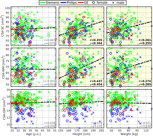
Fig 1
Cross-sectional area of spinal cord white matter correlates with body height and weight. CSA, cross-sectional area; SC, spinal cord; WM, white matter; GM, gray matter; r, Pearson correlation coefficient;ย าฯ, Spearman correlation coefficient. All spinal cord measurements were averaged from cervical C3-4 levels. Regression lines (i.e., the dashed black lines) were estimated from all available data points. Plots with statistically significant correlation (pFWEย < 0.05) are highlighted with yellow background, and corresponding r andย าฯย values are highlighted with black bold font.